Imagine trying to breathe through a straw while someone slowly squeezes the other end. For millions of people, this isn't a thought experiment-it's daily life. Most people use the term Chronic Obstructive Pulmonary Disease is a progressive lung disease that makes it increasingly difficult to breathe over time. Also known as COPD, it isn't just one single condition. Instead, it's an umbrella term that usually hides two very different problems happening in the lungs: chronic bronchitis and emphysema.
While they both lead to shortness of breath, they attack your lungs in completely opposite ways. One clogs the pipes; the other destroys the balloons. Understanding which one is driving your symptoms-or if you have a mix of both-is the difference between a generic treatment plan and one that actually helps you get more air. According to data from the New England Journal of Medicine, patients who get therapy targeted to their specific COPD component see about 27% fewer hospitalizations. Let's break down exactly what's happening inside the chest when these two conditions take over.


The Clogged Pipes: What is Chronic Bronchitis?
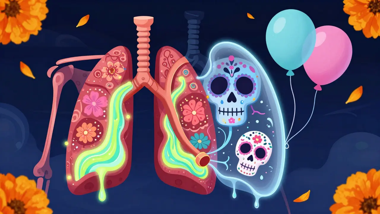
Chronic Bronchitis is a condition characterized by long-term inflammation of the bronchial tubes, leading to a persistent productive cough. Think of your bronchi as the hallways leading to the air sacs in your lungs. In a healthy person, these hallways are clear. In someone with chronic bronchitis, the walls of these hallways swell, and the mucus glands go into overdrive.
Clinically, doctors define this as a cough that produces mucus for at least three months a year, for two years in a row. It's not just a "bad cold" that won't go away. Inside the lungs, there is a massive increase in goblet cells-the cells responsible for producing mucus. In some cases, these glands grow 300% to 500% larger than normal. This leads to the production of 100 to 200 mL of mucus daily, compared to the tiny amount a healthy lung produces. This thick slime blocks the airways, trapping air and making it hard for oxygen to reach the bloodstream.
People with this phenotype are often called "blue bloaters." This isn't a polite term, but a clinical observation. Because the airways are so blocked, oxygen levels in the blood drop (hypoxemia), which can give the skin a bluish tint (cyanosis). The heart often struggles to pump blood through the congested lungs, leading to fluid buildup in the legs, known as peripheral edema.
The Broken Balloons: What is Emphysema?
While bronchitis is about the pipes, Emphysema is a condition where the alveoli, or tiny air sacs in the lungs, are destroyed and lose their elasticity. If the bronchi are hallways, the alveoli are the balloons at the end of the hall where the actual exchange of oxygen and carbon dioxide happens. In emphysema, those balloons pop.
This destruction is usually irreversible. The elastin and collagen fibers that allow your lungs to snap back after a breath are eaten away. This results in a loss of 30% to 50% of the lung's elastic recoil. Instead of many small, efficient balloons, you end up with a few giant, floppy holes. This drastically reduces the surface area available for gas exchange, often cutting oxygen diffusion capacity by 40% to 60% in advanced stages.
These patients are often called "pink puffers." They don't usually turn blue because they compensate by breathing very fast (hyperventilation) to keep their oxygen levels up. However, because the lungs lose their elasticity and trap air, the chest physically changes shape, becoming rounded-a condition known as a barrel chest. Their primary struggle isn't mucus; it's "air hunger," where they feel they can't get enough oxygen despite their lungs being full of trapped, stale air.
Comparing the Two: A Side-by-Side Look
It is easy to confuse the two because both cause breathlessness, but the markers they leave behind are different. If you look at a CT scan, emphysema shows up as low-density "holes" in the lung tissue. Chronic bronchitis looks more like thickened, narrowed walls of the airways.
| Feature | Chronic Bronchitis | Emphysema |
|---|---|---|
| Primary Problem | Mucus & Inflammation | Alveolar Destruction |
| Main Symptom | Productive Cough | Severe Shortness of Breath |
| Physical Sign | Peripheral Edema (Swelling) | Barrel Chest |
| Oxygen Levels | Low (Hypoxemia) | Often maintained via fast breathing |
| Lung Function (DLCO) | Usually Normal | Significantly Reduced |
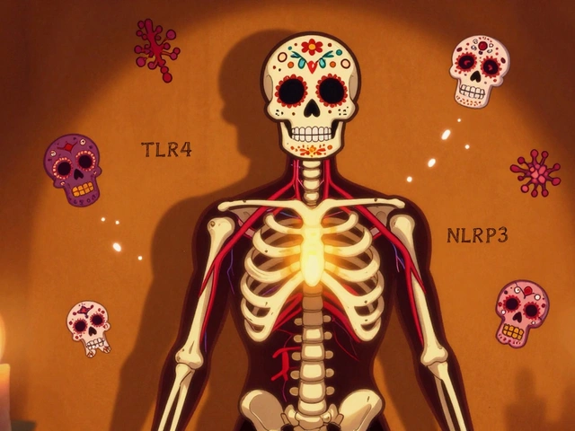
The Overlap: Why the Dichotomy is Often Wrong
For decades, the medical world split patients into "pink puffers" and "blue bloaters." But in the real world, human bodies are rarely that neat. Experts like Dr. Bartolome Celli from Harvard Medical School have pointed out that this split is an oversimplification. In fact, the SPIROMICS study revealed that roughly 85% of severe COPD patients actually show features of both conditions.
You might have the alveolar destruction of emphysema in your upper lobes, while your lower lobes are struggling with the mucus production of chronic bronchitis. This overlap is why a one-size-fits-all approach to treatment often fails. A patient might be using an inhaler to open their airways (which helps the bronchitis part) but still feel suffocated because their lung tissue is destroyed (the emphysema part).
Tailoring the Treatment: Which Approach Works?
Because the pathology is different, the solutions must be different. If you treat emphysema like bronchitis, you might waste time on treatments that don't address the actual structural damage.
For those where Chronic Bronchitis dominates, the goal is mucus management. Mucolytics like carbocisteine are often used to thin the mucus, and some patients find relief through hypertonic saline nebulization. Medications like roflumilast can be a lifesaver for those who have two or more exacerbations per year, reducing those flare-ups by about 17%.
For those with dominant emphysema, the focus shifts to lung volume and oxygenation. Since the air is trapped, some patients undergo lung volume reduction surgery or endobronchial valve placement. These procedures essentially remove the most damaged parts of the lung, allowing the healthier parts to expand and work better. For a small percentage of people, emphysema is caused by alpha-1 antitrypsin deficiency, a genetic condition. These patients require specific augmentation therapy-weekly injections of the missing protein-to stop the lungs from breaking down further.
Practical Steps for Management and Diagnosis
If you or a loved one are struggling with breathlessness, a standard "breathing test" (spirometry) is the first step, but it isn't the whole story. To really distinguish between these components, you need a few specific checks:
- DLCO Test: This measures how well gas moves from your lungs into your blood. If this value is below 60% of what's expected, it's a strong signal that emphysema is the primary driver.
- High-Resolution CT Scan: This allows doctors to see the "holes" of emphysema or the thickened walls of bronchitis.
- The 6-Minute Walk Test: This is a great functional check. Emphysema patients often see their oxygen levels drop sharply within the first two minutes of walking, while bronchitis patients might stop simply because they are too exhausted to keep going.
Beyond the clinic, daily management varies. Those with bronchitis often need 20-30 minutes of chest physiotherapy to clear mucus. Those with emphysema might rely more on portable oxygen concentrators to maintain their mobility. The key is to work with a pulmonologist who looks at your specific phenotype rather than just treating "COPD" as a general label.
Can I have both chronic bronchitis and emphysema at the same time?
Yes, and it's actually very common. Most people with severe COPD have a mixture of both. You might have inflammation and mucus in your airways (bronchitis) while also having damaged air sacs (emphysema). This is why doctors look for a "dominant" phenotype to decide which treatment should be the priority.
Is emphysema curable?
Unfortunately, the destruction of the alveoli in emphysema is irreversible. You cannot "regrow" the air sacs once they are gone. However, treatments like lung volume reduction surgery, pulmonary rehabilitation, and medications can significantly improve your quality of life and slow the progression of the disease.
What is the difference between a "pink puffer" and a "blue bloater"?
"Pink puffers" usually have dominant emphysema; they breathe rapidly to maintain oxygen levels, keeping their skin pink, but often develop a barrel chest. "Blue bloaters" usually have dominant chronic bronchitis; their airways are so clogged that oxygen levels drop, giving them a bluish tint and often causing fluid buildup (edema) in their legs.
How does alpha-1 antitrypsin deficiency cause emphysema?
Alpha-1 antitrypsin is a protein that protects the lungs from being broken down by enzymes. If you are genetically deficient in this protein, your lungs essentially begin to digest themselves, leading to emphysema even if you have never smoked.
Why are LAMA/LABA combinations preferred for some COPD patients?
LAMA (Long-Acting Muscarinic Antagonists) and LABA (Long-Acting Beta-Agonists) are bronchodilators that keep the airways open. They are often preferred over corticosteroids for those with a chronic bronchitis phenotype because some steroids can actually increase the risk of pneumonia in those specific patients.


There are 12 Comments
Del Bourne
Pulmonary rehabilitation is such a game-changer for these patients. It doesn't just focus on the lungs, but helps the rest of the body become more efficient with the oxygen it actually gets. I've seen people regain a huge amount of independence just by learning the right breathing techniques and strengthening their peripheral muscles.
jack hunter
reallly just a way to get ppl on expensive meds... like who cares if you're a puffer or a bloater if the system is just desgined to bleed you dry anyway
Victoria Gregory
It's so important to remember the human side of this!!! 🌸 Breathing is the most basic thing we do, and losing that must be absolutely terrifying. We really need to lead with compassion and love when helping people navigate these diagnoses!!! ❤️✨
Benjamin cusden
The distinction between the two is elementary, though the author's attempt to simplify it for the masses is quaint. Most clinicians would agree that the DLCO test is the only truly definitive metric here, making the 'walk test' more of a qualitative observation than a quantitative diagnostic tool.
dwight koyner
For those managing a dominant chronic bronchitis phenotype, please be mindful of the importance of hydration. Adequate water intake helps thin the mucus, making it significantly easier to clear the airways during chest physiotherapy sessions.
Kathleen Painter
I've always felt that we should look at the intersection of environment and genetics more deeply because while the alpha-1 deficiency is a clear genetic marker, so many people are just products of the industrial areas they grew up in, and it's kind of a tragedy that we treat the symptom without always acknowledging the systemic failure of air quality regulations that let this happen to thousands of people over decades of their lives without any real warning from the authorities.
Ethan Davis
The pharma companies love these 'phenotypes' because it lets them market five different inhalers for the same disease. Just follow the money.
Jamar Taylor
Keep pushing forward everyone! Even if the damage is irreversible, you can still improve your quality of life through consistency and a positive mindset!
Stephen Luce
My dad went through the 'pink puffer' stage for years. It's heartbreaking to watch someone fight for every single breath even when they look okay on the outside.
Nikhil Bhatia
Too much medical jargon.
Laurie Iten
existence is just a long breath we are waiting to let go of i guess
Timothy Burroughs
typical weak lungs result from a weak will to live and total lack of discipline in how we treat our bodies in this godforsaken country where everyone just wants a pill to fix their laziness and avoid the hard work of true wellness
Write a comment
Your email address will not be published. Required fields are marked *