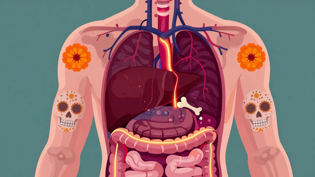
Imagine a blood clot forming in the main highway that carries nutrient-rich blood from your intestines to your liver. This is Portal Vein Thrombosis (PVT), defined as the partial or complete obstruction of the portal venous system by thrombus formation. It’s not just a minor blockage; it can lead to life-threatening complications like intestinal ischemia or worsen existing liver disease. But here is the good news: if caught early, treatment works remarkably well. Early anticoagulation leads to an 85% five-year survival rate. The key? Knowing how to spot it and understanding when-and how-to use blood thinners safely.
What Is Portal Vein Thrombosis?
PVT happens when a clot blocks the portal vein. This vein is crucial because it drains blood from the spleen, pancreas, and gastrointestinal tract into the liver. When this flow stops, pressure builds up behind the clot, leading to portal hypertension.
We classify PVT based on timing:
- Acute PVT: Symptoms appear within two weeks of clot formation. These cases often have a better prognosis because the vessel walls haven’t had time to scar over completely.
- Chronic PVT: The clot has been there for more than six weeks. Over time, the body tries to bypass the blockage by creating new, smaller collateral vessels-a process called cavernous transformation. This makes recanalization (reopening the vein) much harder.
The condition was first described by Rudolf Virchow in 1868. He identified three factors that contribute to clotting: hypercoagulability (blood clots too easily), endothelial injury (damage to the blood vessel lining), and stasis (slow blood flow). Today, we know that about 25-30% of non-cirrhotic PVT cases are linked to inherited or acquired thrombophilias (clotting disorders).
How Doctors Diagnose PVT
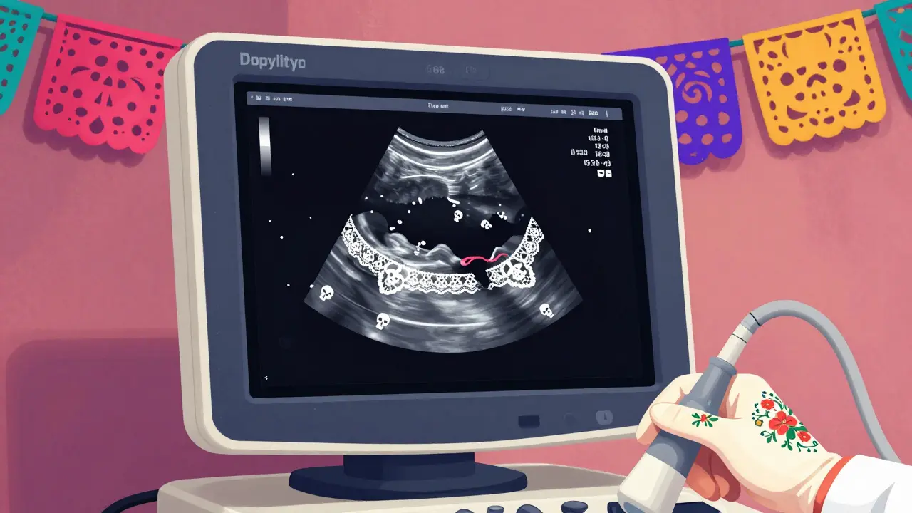
You can’t diagnose PVT with a simple blood test. You need imaging. The gold standard for initial detection is Doppler ultrasonography. It’s non-invasive, widely available, and detects portal vein abnormalities with 89-94% sensitivity.
During an ultrasound, doctors look for specific signs:
- Absence of flow: No blood moving through the portal vein.
- Echogenic material: Visible clot inside the vein.
- Cavernous transformation: A network of tiny collateral vessels replacing the main vein, indicating chronicity.
If the ultrasound is unclear, doctors move to CT portography or MRI portography. These provide detailed maps of the clot’s extent. They classify the occlusion severity:
- Minimally occlusive: Less than 50% lumen obstruction.
- Partially occlusive: 50-99% obstruction.
- Completely occlusive: 100% blockage.
Why does classification matter? Because it dictates treatment urgency. Complete occlusions in acute settings carry a higher risk of bowel infarction, requiring immediate intervention.
Anticoagulation: The Core Treatment
The primary goal of treating PVT is recanalization-getting the blood flowing again. We achieve this through anticoagulation therapy. Current guidelines from the American Association for the Study of Liver Diseases (AASLD) and European Association for the Study of the Liver (EASL) recommend anticoagulation for most acute cases, regardless of whether the patient has cirrhosis, provided bleeding risks are managed.
Timing is everything. Studies show that starting anticoagulation within six months of diagnosis yields a 65-75% complete resolution rate. If you wait longer, that rate drops to 16-35%. Here is how different agents compare:
| Agent Type | Common Drugs | Recanalization Rate (Non-Cirrhotic) | Key Considerations |
|---|---|---|---|
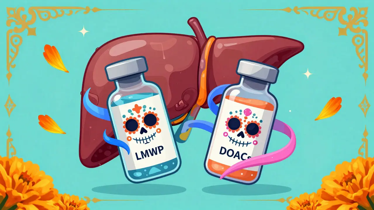
| Low Molecular Weight Heparin (LMWH) | Enoxaparin | 55-65% | Preferred for initial treatment; requires injection; stable control in cirrhotics. |
| Vitamin K Antagonists (VKAs) | Warfarin | 40-50% | Requires frequent INR monitoring (target 2.0-3.0); less effective in advanced liver disease. |
| Direct Oral Anticoagulants (DOACs) | Rivaroxaban, Apixaban | 65-75% | Oral administration; superior resolution rates in non-cirrhotics; caution in Child-Pugh C. |
For non-cirrhotic patients, DOACs like rivaroxaban (20 mg daily) or apixaban (5 mg twice daily) are increasingly popular due to their convenience and high efficacy. However, in patients with cirrhosis, LMWH remains the preferred choice for many specialists because it offers more predictable anticoagulation levels without relying on liver metabolism.
Treating PVT in Cirrhotic Patients
Managing PVT in someone with cirrhosis is tricky. These patients already have fragile blood vessels and a higher risk of bleeding. Major bleeding complications occur in 5-12% of cirrhotic patients on anticoagulation, compared to 2-5% in those without cirrhosis. Variceal bleeding accounts for 60-70% of these events.
So, do we treat them? Yes, but carefully. The 2023 EASL guidelines recommend endoscopic variceal screening before starting any blood thinner. If large varices are found, doctors perform band ligation to secure them. One study from UCLA Medical Center showed that performing endoscopic variceal ligation before initiating anticoagulation reduced major bleeding events from 15% to 4%.
Anticoagulation is generally contraindicated in:
- Patients with recent variceal hemorrhage (within 30 days).
- Those with uncontrolled ascites.
- Patients with Child-Pugh Class C cirrhosis (severe liver dysfunction).
For those with compensated cirrhosis (Child-Pugh A or B), the benefits of preventing clot extension usually outweigh the bleeding risks. The 2024 AASLD guidance update even expanded DOAC indications to include some Child-Pugh B7 patients, based on recent trial data showing comparable safety to LMWH.
When Blood Thinners Aren't Enough
Sometimes, anticoagulation fails. The clot doesn’t dissolve, or it grows despite treatment. In these cases, interventional procedures become necessary.
Transjugular Intrahepatic Portosystemic Shunt (TIPS) is a common second-line option. Radiologists create a channel between the portal vein and hepatic vein using a stent. This bypasses the clot and reduces pressure. TIPS has a 70-80% technical success rate but carries a 15-25% complication rate, including hepatic encephalopathy (brain fog caused by toxin buildup).
Another option is percutaneous mechanical thrombectomy, where doctors physically remove the clot using catheters. This shows 60-75% immediate recanalization rates but is limited to specialized centers. Surgical shunts are rarely used today due to high morbidity.
Duration of Therapy and Follow-Up
How long do you stay on blood thinners? It depends on why the clot formed.
- Provoked PVT: If the clot was triggered by a temporary factor (like surgery or infection), treatment lasts at least 6 months after the factor resolves.
- Unprovoked/Thrombophilia: If you have an underlying clotting disorder (present in 25-30% of non-cirrhotic cases), lifelong anticoagulation is often indicated.
- Malignancy: Patients with active cancer-related PVT typically require indefinite therapy until the cancer is controlled.
Regular follow-up includes repeat ultrasounds every 3-6 months to check for recanalization. Doctors also monitor liver function tests and platelet counts. For patients on warfarin, INR checks are essential. For those on DOACs, renal function must be monitored since these drugs are cleared by the kidneys.
Emerging Trends and Future Directions
The landscape of PVT treatment is evolving. The FDA approved andexanet alfa in 2023, a reversal agent for factor Xa inhibitors. This addresses a major safety concern: what to do if a patient on a DOAC starts bleeding uncontrollably. Previously, reversing these drugs was difficult.
Research is also focusing on precision medicine. Genetic markers like F5 Leiden and PROTHROMBIN G20210A help predict which patients will respond best to extended anticoagulation. Ongoing trials, such as the 500-patient RCT comparing rivaroxaban versus enoxaparin in cirrhotic PVT, aim to refine our understanding of optimal drug choices. By 2027, targeted thrombolysis may see wider adoption, offering a faster way to dissolve clots in critical cases.
Can Portal Vein Thrombosis resolve on its own?
It is rare for a significant PVT to resolve without treatment. While small, incidental clots might stabilize, untreated PVT often progresses to chronic disease with cavernous transformation. Early anticoagulation is crucial to prevent permanent damage and intestinal ischemia.
Is it safe to take DOACs if I have liver cirrhosis?
Safety depends on the severity of your liver disease. DOACs are generally avoided in Child-Pugh Class C cirrhosis due to high bleeding risks and unpredictable drug metabolism. For compensated cirrhosis (Class A or B), recent guidelines suggest they can be used cautiously, especially after variceal screening and banding. Always consult a hepatologist.
What are the symptoms of acute PVT?
Acute PVT can present with sudden abdominal pain, tenderness, nausea, vomiting, and bloody stools if intestinal ischemia occurs. Some patients may experience fever or worsening ascites. However, many cases are asymptomatic and discovered incidentally during imaging for other conditions.
How long does anticoagulation therapy last for PVT?
The duration varies. For provoked cases (e.g., post-surgery), treatment typically lasts 6 months. For unprovoked cases or those with underlying thrombophilia, lifelong anticoagulation is often recommended to prevent recurrence. Regular imaging monitors progress to determine if therapy can be adjusted.
Does PVT affect liver transplant eligibility?
Historically, PVT complicated transplants, but modern techniques have changed this. Anticoagulation improves post-transplant survival rates (85% one-year survival in treated vs. 65% in untreated). Many centers now list patients with PVT for transplant, sometimes using TIPS or surgical shunts to facilitate the procedure.





There are 9 Comments
Joseph Teichman
Good info. Helps me understand my dad's condition better. Keep it simple like this.
Grace Gayle McMullen
Hey there, just wanted to say that the section on DOACs vs LMWH is super helpful for folks trying to navigate these choices with their doctors. I've seen so many people get confused about why warfarin isn't always the first pick anymore. It's wild how much guidelines have shifted in just the last few years. Also, the bit about endoscopic screening before starting blood thinners in cirrhotics is a lifesaver literally. So many people skip that step and then deal with variceal bleeding which is a nightmare. Hope this helps someone out there who's scared of the diagnosis.
Angela Niculescu
I honestly think the article oversimplifies the risk of DOACs in liver patients. Saying they are 'increasingly popular' ignores the fact that we still don't have enough long term data on safety in Child-Pugh B patients. It feels like marketing hype rather than solid evidence based medicine. The recanalization rates look good on paper but what about the bleeding risks that aren't fully captured in those short term studies? People need to be more skeptical of these new trends.
Victoria Mangiapane
Ugh another medical post making everything sound easy. Like yeah sure just take a pill and you'll be fine. But what about the side effects? What about the cost? This reads like a brochure not real life advice. Typical.
Jonhnnie john13
The logic here is flawed. You present anticoagulation as a silver bullet but ignore the high failure rate in chronic cases. Cavernous transformation makes recanalization nearly impossible yet the tone suggests otherwise. This is dangerous misinformation disguised as education. Stop selling hope where none exists.
Anthony Padilla
Hey everyone, i really appreciate this detailed breakdown especially for our global community dealing with different healthcare systems. In india we often see delayed diagnosis due to lack of access to doppler ultrasound early on. It would be great if more resources were translated or made available locally. Also wondering if anyone has experience with traditional therapies alongside western medicine? Just curious about holistic approaches that might support liver health during treatment.
Elizabeth Fandry
One must consider the existential weight of such vascular obstructions 🧠💉. The body’s attempt at cavernous transformation is a poetic yet tragic testament to human resilience against entropy. While the clinical data on DOACs is compelling, one cannot ignore the philosophical implications of relying on synthetic agents to correct nature’s errors. Are we truly healing or merely delaying the inevitable collapse of our biological infrastructure? The FDA’s approval of andexanet alfa is a small victory in the grand theater of mortality, but let us not mistake convenience for wisdom. True understanding requires grappling with the fragility of our own veins 🩸✨.
Madeline Petes
Wow this is actually really empowering! I was so scared when my friend got diagnosed but reading that 85% survival rate gives me so much hope. The part about TIPS being an option if meds dont work is also reassuring. We just gotta stay positive and follow the doctors orders right? 💪🌟
Ramanath Rao
This article is pathetic. It fails to address the stark disparities in PVT management between developed and developing nations. While you discuss DOACs and TIPS, millions lack basic diagnostic imaging. Your focus on 'convenience' is insulting to those who struggle to afford enoxaparin injections. Wake up to the reality of global health inequity instead of preaching to the choir in wealthy countries. This is not just a medical issue it is a systemic failure.
Write a comment
Your email address will not be published. Required fields are marked *